First impressions of OMAX M837ZL series microscope
The microscope I bought arrived, and I spent a day getting familiar with it and the lenses. I was generally pleased, but ended up buying a bunch of new objectives and condensers to see if I could get better images. This post is regarding the initial purchase except the images at the end featuring the darkfield condenser.
This is part of a series:
- I bought a microscope
- Seeking OMAX M837ZL series microscope manual
- First impressions of OMAX M837ZL series microscope
- OMAX software and micrometer sample images
- First tardigrade sighting!
Upon opening the box, I found the sheet instructing to download the manual as noted by the now removed Amazon review.

Unboxing the OMAX M837ZL microscope

Unboxing the OMAX M837ZL microscope
I wrote in “Seeking OMAX M837ZL series microscope manual” about how I initially couldn’t access the manual since I typed the URL incorrectly and then tried to find the microscope product page, which turned into a bit of a project in itself. Once I found the microscope product web page, I realized there is no link to relevant manuals or software downloads! I continued unpacking.

Unboxing the OMAX M837ZL microscope

Unboxing the OMAX M837ZL microscope

Unboxing the OMAX M837ZL microscope

The image sensor of the OMAX 14MP USB3 microscope camera

Unboxing the OMAX M837ZL microscope
I was amused to find the CD supposedly containing the software and manual was damaged.

My OMAX M837ZL microscope came with a broken CD, but luckily I was still able to get the files off.

My OMAX M837ZL microscope came with a broken CD, but luckily I was still able to get the files off.
I described finding the software and my initial testing in “OMAX software and micrometer sample images“.
Trying it out
I initially started looking for tardigrades and documented that in “First tardigrade sighting!“, but also did some cheek cell samples below.
Here’s my desk setup. It’s a good thing I mounted the monitor on the wall so I could find more devices to fill my desk space!

First test of the OMAX M837ZL microscope, looking at some local moss associated cells.
The color isn’t great on this next image, and I wrote more about the white balance problems with the OMAX 14MP USB 3.0 camera in my micrometer sample images entry. I had better luck with the Nikon attachment, but I didn’t have that yet when I made this image.
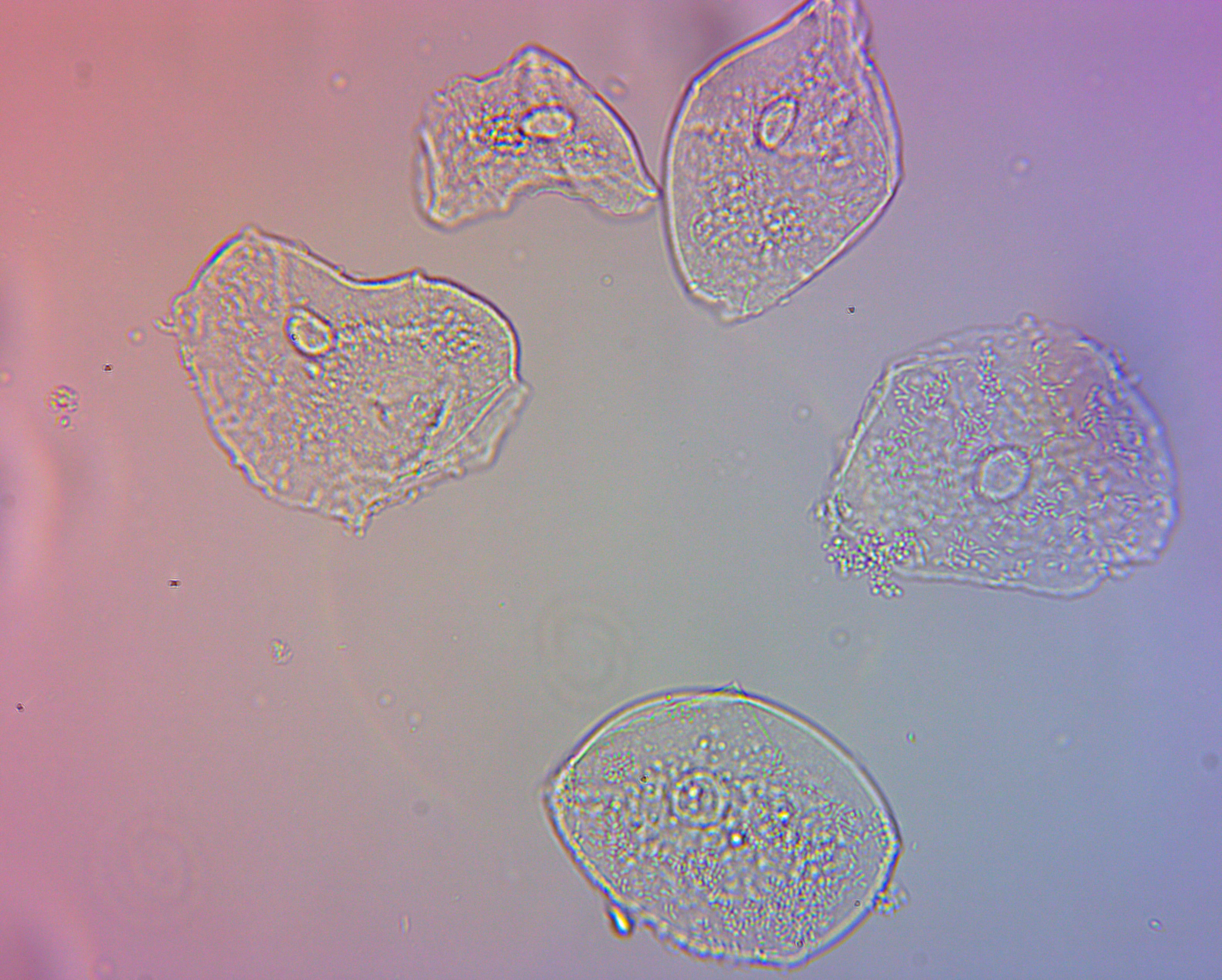
Human cheek cells viewed through an OMAX achromatic 40X NA0.65 160/0.17 objective lit through an OMAX dry brightfield Abbe NA1.25 condenser captured by an OMAX 14.0MP USB3.0 camera with 0.5X adapter
The next two images are the same cells using my new darkfield condenser. This was my first time using a darkfield condenser, so I may not have adjusted things properly. I experimented with different white balance settings, hence the blue and green images. You can see different features of the cells are visible with the different white balance settings.
I am pretty sure this shows dozens of small bacterial cells covering the outsides of the cheek cells. Pretty fascinating!
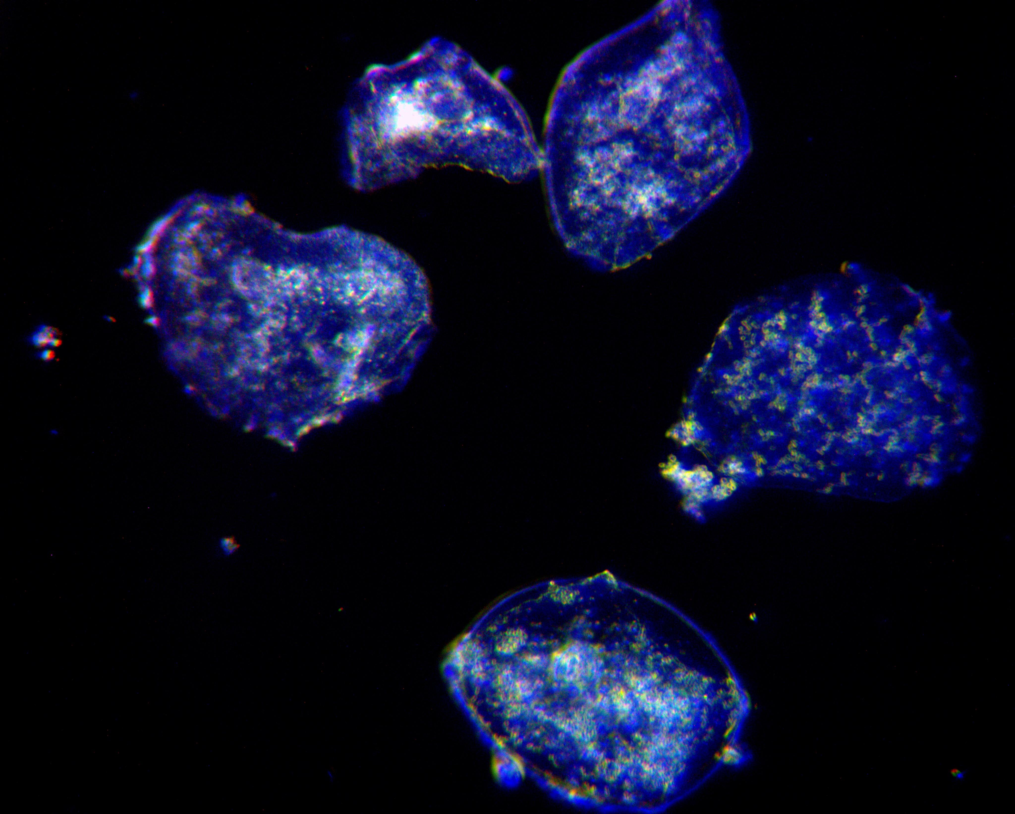
Human cheek cells viewed through an OMAX achromatic 40X NA0.65 160/0.17 objective lit through an OMAX dry darkfield NA0.7-0.9 condenser captured by an OMAX 14.0MP USB3.0 camera with 0.5X adapter
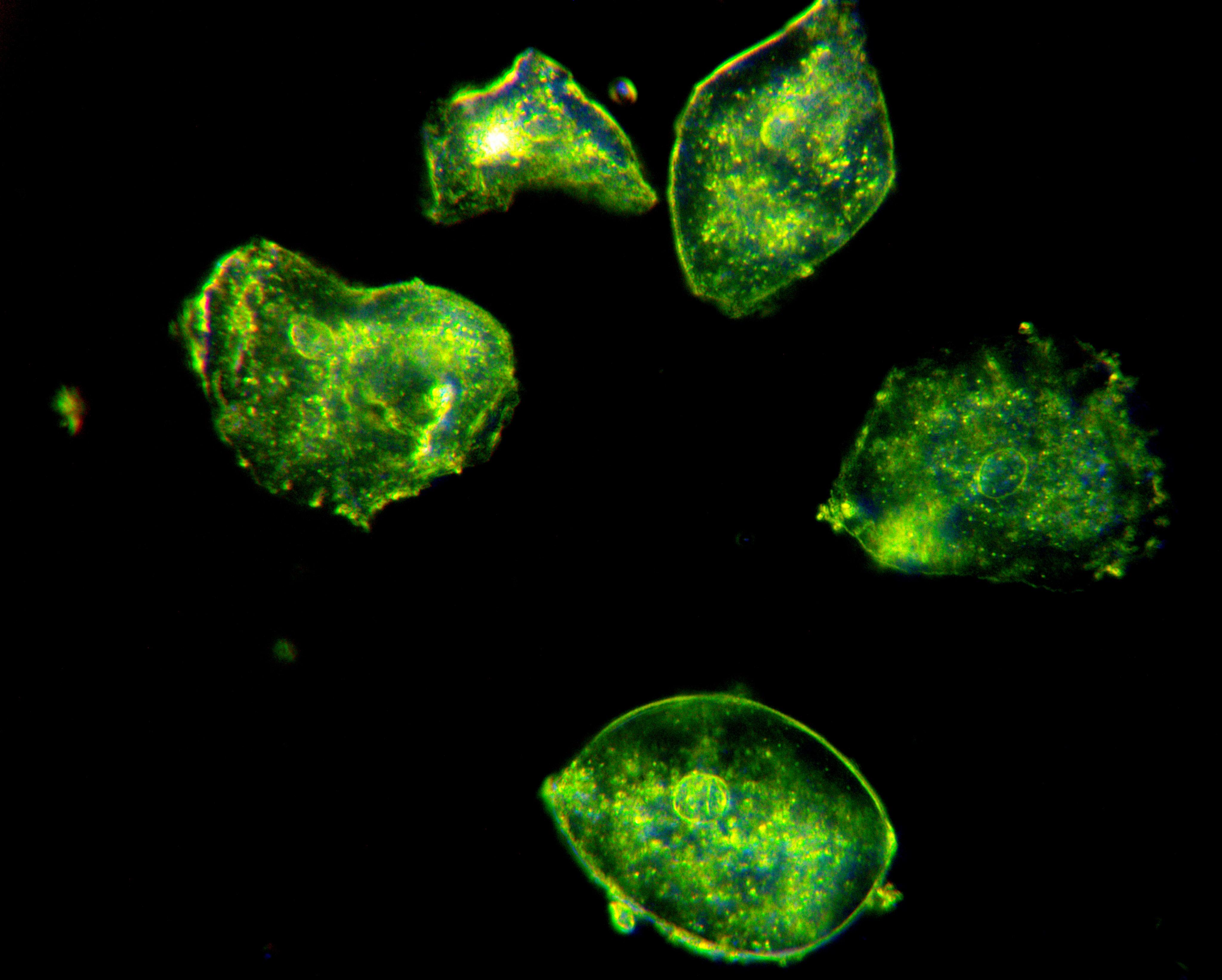
Human cheek cells viewed through an OMAX achromatic 40X NA0.65 160/0.17 objective lit through an OMAX dry darkfield NA0.7-0.9 condenser captured by an OMAX 14.0MP USB3.0 camera with 0.5X adapter
I tried looking at the cells a day or two later when I received my first “plan” objective lens, which should have allowed me to get more in focus at once, but it seems the cells dried and shriveled up, even under the cover slip and sealed with nail polish. I’m not sure if this is just drying, or they reacted due to the nail polish, or something else.
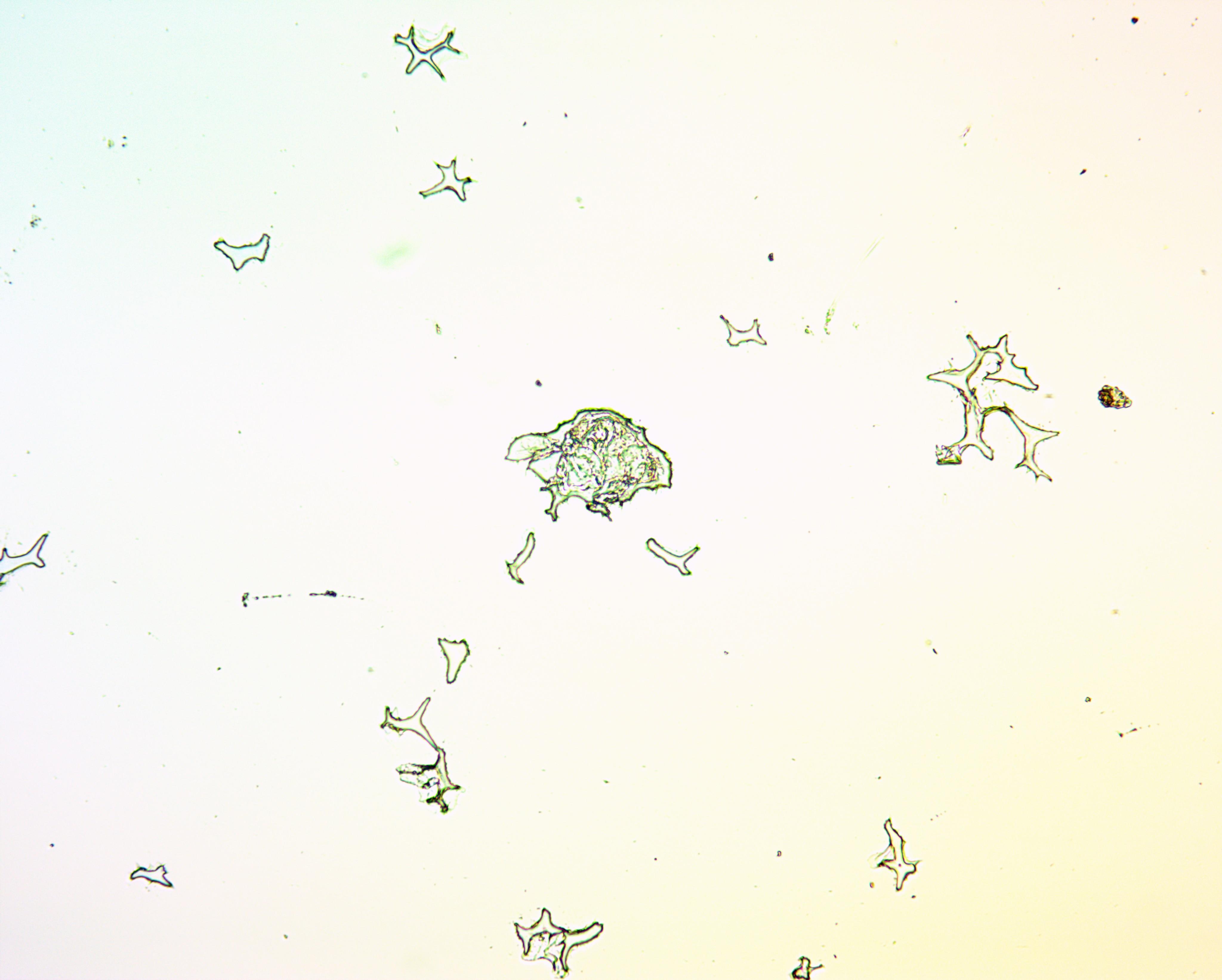
Dried, shriveled human cheek cells on a slide left out for a day viewed through an OMAX achromatic 10X NA0.25 160/0.17 objective lit through an OMAX dry brightfield Abbe NA1.25 condenser captured by an OMAX 14.0MP USB3.0 camera with 0.5X adapter
Impressions
I guess this section was supposed to be the whole point of this entry, where I would write almost a review of the microscope after using it for a few hours. I’m pretty tired now after putting all the rest of these entries together, so I may have to come back to this.
I’ll just say for now:
Overall I enjoyed using the scope, and it felt fairly familiar from my days in the biochemistry lab during college. I did feel quite limited by the objectives, and will do further comparisons of them and other lenses in my micrometer sample images entry. The 4X lens was fine, but the 10X and 40X showed the strong red and blue coloration on either side of the image. I later found this is apparently an artifact of the OMAX camera, and I realize it didn’t show up in the eyepieces either. I’ll have to withhold judgment on that for now.
I found I needed to cover parts of the microscope head with electrical tape to block light entering where I don’t think it was supposed to. Before doing this, I saw an image of my window and curtains floating around the sample.
Using the stage and focus controls was straightforward. I would have liked it to be easier to make finer movements with the stage for higher magnifications, but this scope didn’t perform any worse than other scopes I’ve used. I just had to tighten my hand like a large gear and move with my elbows to achieve a smooth, small motion, instead of the jerks I would have gotten if I just grabbed it and turned with my fingers.
The 3 watt LED light was more than sufficient for brightfield work, but when I used the darkfield condenser, especially the oil one I bought for higher NAs, the LED light was not bright enough. I compensated by taking photos with my Nikon D800 with high ISO, but I wasn’t able to see much in the eyepieces at 63X or 100X and darkfield.
I’ll write more later perhaps!